 Favoritos de f-sundw


Fotos / Sonidos

Autor
seastungDescripción
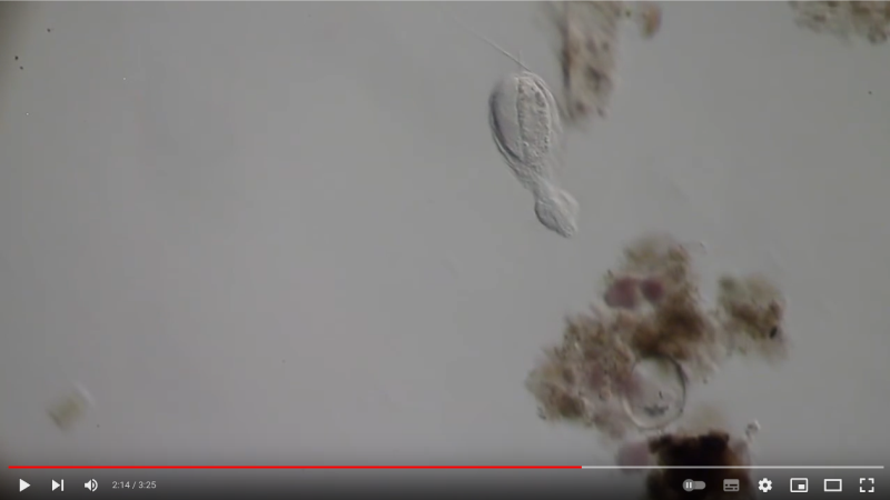
this one in a capsule that is seen in some of the images, billowing about.. any ideas?

Fotos / Sonidos

Qué
Género CoryneAutor
luca_dtDescripción
Quite common. Display positive phototaxis at night.


Fotos / Sonidos

Qué
Sarsia princepsAutor
cemillsDescripción
Caught in the wild and photographed nearby in an aquarium. Bell is 20–25 mm tall.
Note tall, conical aspect of the bell and the gonad extends from nearly the top of the manubrium to the mouth (just a very short section at the top of the long manubrium is without gonad).


Fotos / Sonidos

Qué
Género PythiumAutor
crseaquistDescripción
Taken from the top 3cm of dry soil next to a playa on 2023-08-27.
For video see: https://youtu.be/Nrzh5UoG9gE


Fotos / Sonidos

Qué
Mariposa Búho de Ojos Cafés Centroamericana (Dynastor darius ssp. stygianus)Autor
magazhuDescripción
Dia 82, un individuo.
Day 82, one individual.
Relacionado con // Related to:
https://www.inaturalist.org/observations/2667158
https://www.inaturalist.org/observations/2667199
https://www.inaturalist.org/observations/2667420

Fotos / Sonidos

Qué
Hongos (Reino Fungi)Autor
gustaf1Descripción
On gorse. Conidiaphores emerging from black embedded sporodochia. Conidiaphores septate, pigmented, erect, and occasionally branching. Forming conidia in regular intervals up the condiaphore. Conidia hyaline, oval, 7.36-9.43×4.6-5.75 uM, avg 8.58×4.93 n=7
Fotos / Sonidos

Autor
dms1535Descripción
Haltidytes crassus was a relatively common species at the time of its first description. Since then - probably due to the influence of industrial agriculture - this pretty gastrotrich has become very rare.
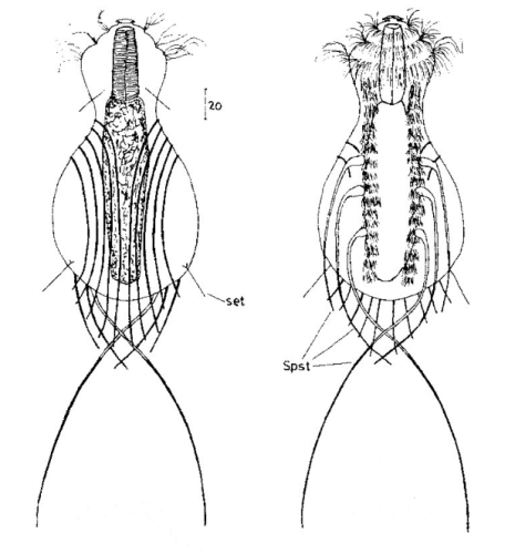
The main characteristic of H. crassus are the five dorsal and three ventral “jumping spines”, with which the animal catapults itself out of the danger zone when threatened.
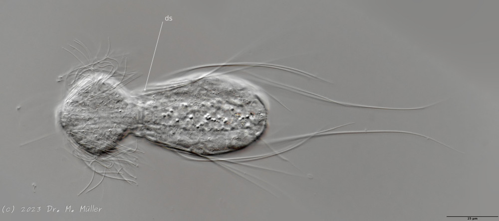 Dorsal (juvenile animal); ds: dorsal spines (5 pairs).
The long, caudal and hair-shaped last ventral pair of spines, which far overhang the body and cross each other in resting position, is conspicuous. Three ciliary rings on the head are used for swimming locomotion, while the ventral ciliary bands typical of gastrotrichs are used for gliding over the substrate.
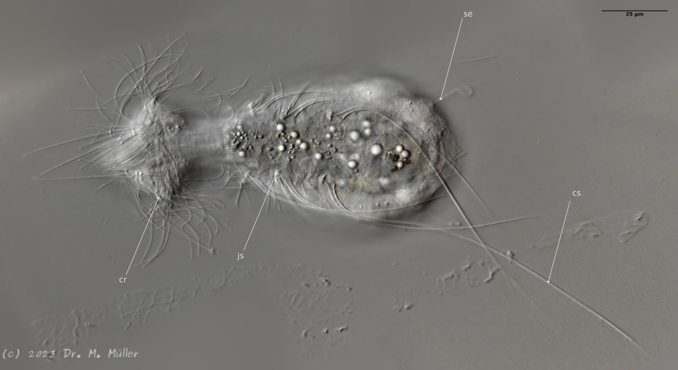
Ventral; cr: 3 ciliary rings for locomotion; js: jump spines; cs: long caudal spines; se: seta on papillae.
The posterior palps - as in all members of the genus Hatidytes - are on distinct papillae.
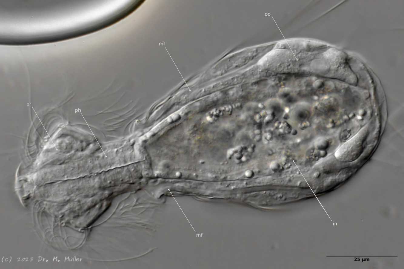
Median; br: “brain”; ph: pharynx; oo: mature ovum; in: “intestine”; mf: muscle fibers that move the jumping spines.
The internal anatomy has some peculiarities. For example, distinct muscle fibers can be seen that couple to the attachment sites of the jumping spines. Adhesive glands and the associated adhesive tubes are not present in this genus.
The typical movement and behavior of the animals can be seen in the following YouTube video:
Movie about Haltidytes crassus


Fotos / Sonidos

Autor
claudenDescripción
Discovered when cleaning a tank at institute, presumably came in from the seawater intake.
Fotos / Sonidos

Autor
kallamperoLugar
Permanent Plots, Reserva Los Cedros, Intag, Cotacachi Canton, Imbabura Province, Ecuador (Google, OSM)Descripción
Substrate: unk. Xylaria sp.
Habitat: Northwest Andean montane forest (NT0145)
Collectors: D. Newman & R. Vandegrift
Collection #: RLC1340B
Photomicrography and molecular data forthcoming
Fotos / Sonidos

Autor
joshmcginnisDescripción
Isolated from the surface of Annulohypoxylon growing on a dead log alongside Tremella fuciformis (https://www.inaturalist.org/observations/101722719).


Fotos / Sonidos

Autor
gustaf1Descripción
Fungi on Veronica sp. Asexual spores septate with one gluttule per cell, spores 11.5-18.5×6.21-8.05 uM, avg 17.6×6.54 n=7.


Fotos / Sonidos

Qué
Hongos (Reino Fungi)Autor
gustaf1Descripción
On bird bone. Conidia oval to round, 6.9-4.6×4.6-3.45 uM, avrg: 5.56×4.22 n=6
Fotos / Sonidos

Qué
Hongos de Saco, Levaduras Y Parientes (Filo Ascomycota)Autor
gustaf1Descripción
On ash key. Very easily scraped off. It seems to me that it begins immersed in the wing and then "bursts" out, as indicating by the orange pustules that are visible in picture 4. Anamorph orange and unstructured, just a spore mass. Spores 17.25-13.8×6.9-5.75, avrg 6.77×2.73 n=7.
Fotos / Sonidos

Autor
gustaf1Descripción
Anamorph of fungi growing on incubating wood sample with Dendrostilbella prasinula. Conidiophore is septate. Conidia oval tapering at one end and 2-3 septate, length 18.86-21.85 uM, width 8.05-9.66 uM.
Fotos / Sonidos

Autor
leonardomondacalaraDescripción
23 m de profundidad, sustrato rocoso, múltiples ejemplares.
Valva dorsal y ventral.
Fotos / Sonidos

Autor
luca_dtDescripción
A few washing ashore.
This genus due to exumbrella cnidocyst tracks and radial canal lengths (from NIWA memoir 113). But not H. prolifer due to tentacle count and shape.
I wonder if these are the common solitary hydroids we get around the coast.
~3mm tall


Fotos / Sonidos

Qué
Gran Argonauta del Pacífico (Argonauta argo)Autor
invertebratistDescripción
79.5 mm.
Jill Griffiths Collection.
''Approaching fullmoon. Stormy seas. Within 2hrs lowtide and yet running very high'' From the label.
This is the first (and currently only one) accurate observation of Argonauta argo from NZ on Inaturalist.

Fotos / Sonidos

Autor
invertebratistDescripción
Native bush, in wet plant litter among fallen Nikau fronds.
Body length 37 mm.(antennae not included)
Probably unnamed. I would call it Peripatoides aff. indica or P. aff. aurorbis.
Absolutely splendid & my very best find in 2022.
I am still crazy excited by this. The colour was literally velvet.
I released the individual after photography.
My post of this Velvet worm achieved 100k likes and 11 million impressions on my twitter(url)!


Fotos / Sonidos

Qué
Haya Común (Fagus sylvatica)Autor
kbs_64Descripción
Tatsächlich! Eine Albino-Buche!


Fotos / Sonidos

Autor
aleshirokikh32Fecha
Septiembre 15, 2014Descripción
These Myxomycetes form a community that was located on a fallen aspen trunk (Pópulus trémula).

Fotos / Sonidos

Autor
zihaowangDescripción
200x and 800x magnifications. Fruiting bodies seemed to only form on the abaxial leaf surface of Quercus bicolor.
Fotos / Sonidos

Autor
leeormsby67Descripción
Found on a very rotten branch, probably Tawa. My thoughts on ID are that it looks Mycena-like? Awaiting microscopy by Shirley Kerr. Cap surface on mature specimens was very sticky/tacky to the touch.

Fotos / Sonidos

Autor
ivijayanandDescripción
Fungi on an insect / spider carcass
Location: Makunda Christian Hospital, Karimganj District, Assam
Date: 1st October 2012
Equipment: Nikon D300s with Nikkor AF 28-105mm lens
Fotos / Sonidos

Autor
calliebroaddusDescripción
Over 5' long, found crossing the road in NW Ecuador at Dracula Reserve, around 2,000m elevation.


Fotos / Sonidos

Autor
alison_pollackDescripción
Substrate is Umbellularia californica (California Bay Laurel) nut.
Fotos / Sonidos

Autor
kallamperoDescripción
Comments: this was the largest of any “bamboosicle” I have ever witnessed in person, dry or fresh. I so wish I were the one to find it in the field, and could have therefore photographed it in situ, but stumbling upon it back in the lab was still a pure delight. the final image shows some last-minute shots of the largest fruiting body — which had the heft and overall gestalt of a chicken pot pie — with my hand an a coin for scale, respectively. this species’ size, locality, and truly shelf-like growth habit all make the ID unmistakable as A. polyporoides. also note the typically long, filiform ascospores ejecting against the black background in the close-up shots.
Originally posted to Mushroom Observer on Apr. 29, 2019.


Fotos / Sonidos

Autor
matthew_connorsDescripción
Nice star-shaped barnacles on the side of the pier


Fotos / Sonidos

Qué
Physarum oblatumAutor
alison_pollackDescripción
Collected by Sittee Aisha, graduate work for Dr. Steven Stephenson, University of Arkansas.
Fotos / Sonidos

Autor
alison_pollackDescripción
Found by @graysquirrel . Thank you Krissa! It is a wonderful find!
Fotos / Sonidos

Autor
millenialmycesDescripción
I’m not confident on the species ID, but looks like Ophiocordyceps. I found it on a Carabidae ground beetle under oak. If you are interested in the specimen, let me know. I have it stored.
Fotos / Sonidos

Autor
cgmayersDescripción
Growing in gallery of Scolytoplatypus fasciatus in Nuxia floribunda. In the microscopic images it is stained with cotton blue; the fungus itself is whitish-clear.
Fotos / Sonidos

Autor
pbertnerDescripción
Presumably a parasitic plant since no leaves were observed and no plants nearby.
Fotos / Sonidos

Qué
Eperetmus typusAutor
cemillsDescripción
Collected in late summer at the surface in Friday Harbor, photographed in an aquarium. Bell diameter was 30 mm. A rare visitor near the end of the season, when there were very few other jellies in the water. See a beached one August 2021 from Homer, Alaska at https://www.inaturalist.org/observations/95334168.


Fotos / Sonidos

Autor
russellcummingDescripción
Jandakot Regional Park, off Anstey Road.
Photos 5 & 6 - Utricularia violacea and U. multifida.
Fotos / Sonidos

Autor
max_colemanDescripción
Naturalised in a wooded stream valley close to the sea. Population of several hundred individual plants. Anthers yellow and filaments swollen. Not mentioned in the county flora or recorded for the vice county in the BSBI database.


